Next in our weekly series of articles, Dr. Danielle Hamilton, a Research Scientist with the Centre for Chromosome Biology, writes about her work “Exploring the Cell” and how understanding how a cell repairs damage to its DNA may lead to the prevention and treatment of cancer.

Every living creature is made up of one or more cells, and humans are no exception. These microscopic structures are the building blocks of our bodies and each is programmed to perform a specific function. Cells of the same type are often found clustered together and communicate with each other to form the tissues and organs that make up a functioning organism.
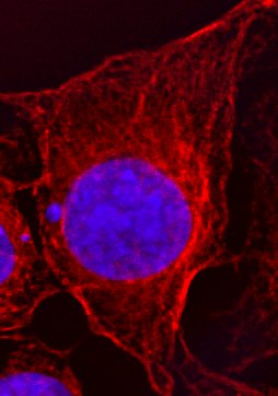
Cells are composed of a liquid known as the cytoplasm enclosed within an outer layer called the plasma membrane. Important structures known as organelles are found within the cytoplasm, and these include the mitochondria (small organelles that produce energy for our cells), ribosomes (responsible for decoding our DNA to produce proteins), and the nucleus (the organelle that contains our DNA).
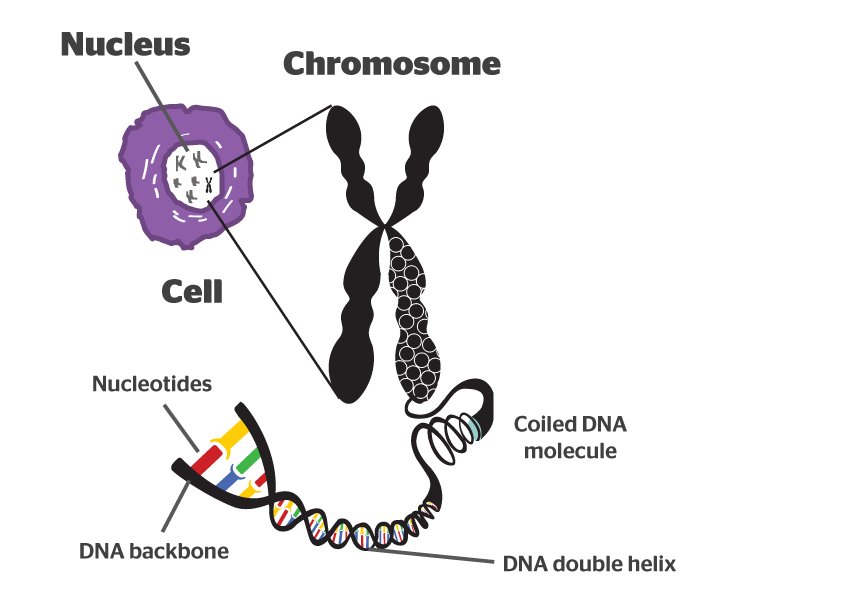
Our research at the Centre for Chromosome Biology in NUI Galway focuses on the DNA within the nucleus. DNA is compacted into structures known as chromosomes, of which each of our cells contains 23 pairs. DNA is similar to a computer code and contains all of the information our cells require to carry out their functions. This code is composed of different combinations or ‘sequences’ of the nucleotides, Adenine (A), Thymine (T), Cytosine (C) and Guanine (G) and can be decoded by a tiny organelle called the ribosome, to produce amino acids, which are the basic units of proteins.
Genes are regions of the genetic code that encode specific proteins and are inherited from our parents. Some genes’ sequences can vary from person to person without any damaging consequences; for example, differences in hair colour arise from variations in the gene encoding the protein melanin. However, some genes are so important to our cells that any changes to their code will result in cell death. For example, an accidental change to a gene that controls cell metabolism can prevent the cell from producing enough energy, eventually leading to its death.
I work in the Genome Stability Laboratory, where we study the genes responsible for repairing and protecting our DNA. Mistakes can be introduced into the genetic code accidentally during normal DNA replication or during cell division.
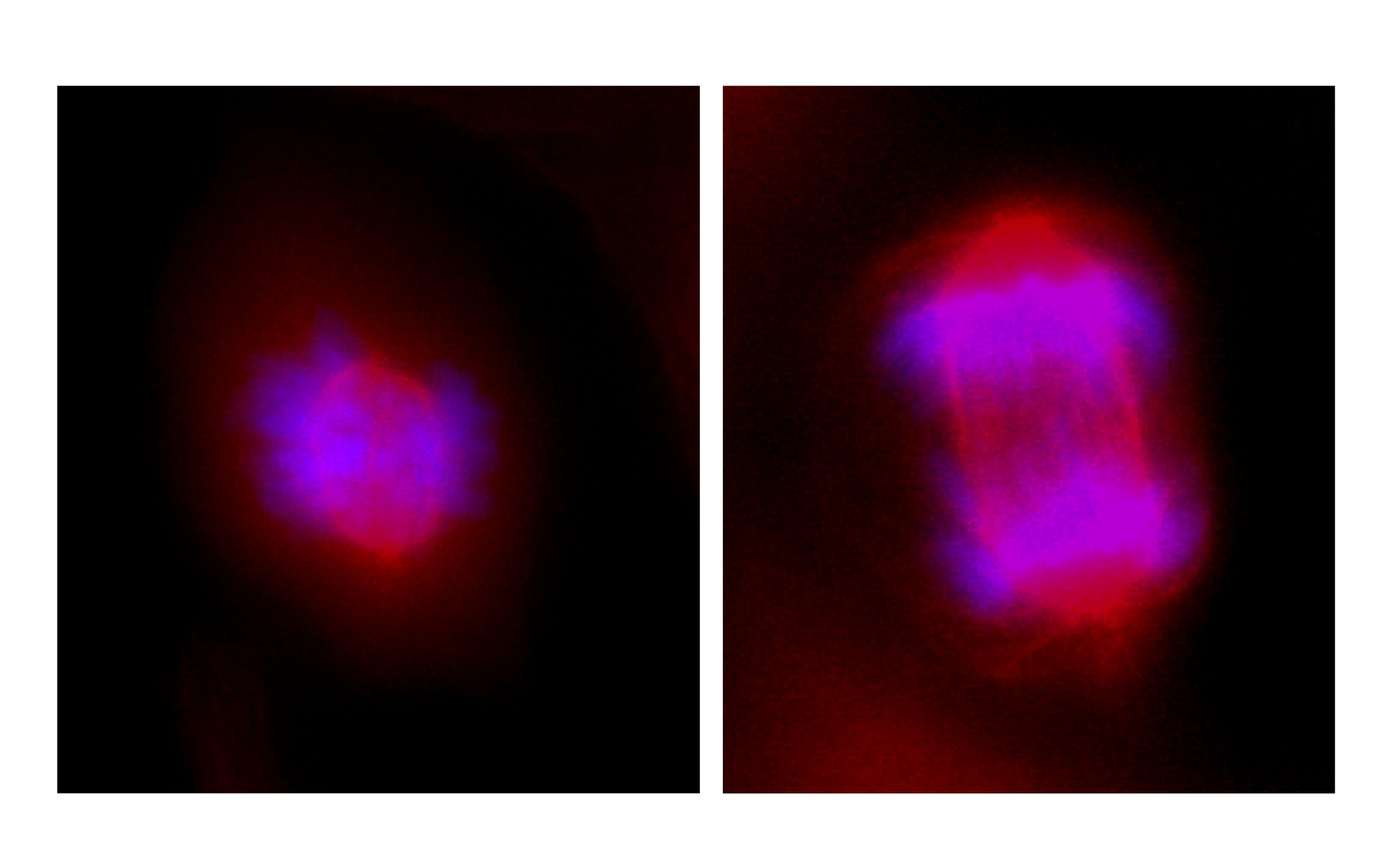
Above is a short time-lapse movie showing cell division filmed by Dr. Emma Harte, Research Scientist, Centre for Chromosome Biology, NUI Galway. The panel on the left shows the DNA inside the cells, while the panel on the right shows the outer membrane surrounding the cell.
External sources such as cigarette smoke, UV radiation from the sun and Ionising Radiation from atomic bombs can also cause damage to our DNA. Fortunately, a process known as the DNA Damage Response is triggered in such a situation and this allows our cells to stop dividing and focus on repairing the damaged DNA. This is particularly important in the prevention of cancer, a disease arising from the accumulation of DNA mutations.
If the DNA damage response is defective, this allows DNA mutations to go undetected and uncorrected, resulting in unusual and unpredictable cells. Not all mutations cause cell death, for example, many cancer cells have mutations in genes regulating cell division that allow them to grow faster. This makes it much easier for them to outgrow their neighbouring cells and form tumours.
My work focuses on uncovering new genes required for the repair of genetic mutations. To do this I use genetic engineering to silence specific genes in human and mouse cells and examine how the cells respond to DNA damage. Discovering new genes required for DNA repair will hopefully lead to a better understanding of cancer development and help us to find new targets for cancer therapy.
If you’d like to find out more about becoming a researcher in the Centre for Chromosome Biology, check out their Recruitment page or if you’re interested in studying Biochemistry in NUI Galway, check out the videos below:

